Combining cryo-EM and mass spectrometry imaging capabilities into a single workflow provides unprecedented spatial and chemical composition detail of biological samples
Electron cryomicroscopy (cryo-EM) is an invaluable tool for determining molecular structures, capable of visualising biological samples with high spatial resolution. However, whilst the technique allows researchers to distinguish shapes and contrast of features in their images, it cannot provide any information regarding the chemical composition of the imaged sample. Conversely, mass spectrometry is a technique which provides compositional information but is unable to give detailed spatial information. Akin to adding colour to a black-and-white photograph, augmenting cryo-EM spatial data with mass spectrometry-derived chemical detail would greatly expand the information scientists can gather. Tanmay Bharat’s group, in the LMB’s Structural Studies Division, has developed a novel workflow for correlated electron microscopy and imaging mass spectrometry which achieves just this.
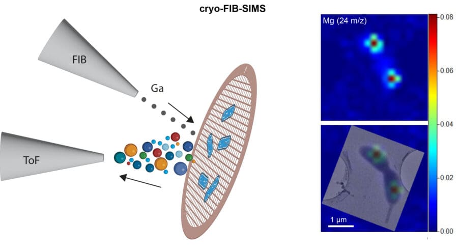
Spearheaded by postdoc Hannah Ochner, the new workflow first captures high resolution structural information using cryo-EM, followed by imaging mass spectrometry of the same sample using a focussed ion beam scanning electron microscope adapted with a time-of-flight mass spectrometer (FIB-SIMS, focussed ion beam secondary ion mass spectrometry), allowing correlation between both types of data. Using this new cryo-EM-FIB-SIMS workflow, the team were able to track compounds at unprecedented resolutions, providing a subcellular map with more information than either of the composite techniques can singularly.
This new workflow is compatible with numerous sample types and can be combined with other imaging methods. Using dual metal and fluorescently tagged bacteria prepared by Buse Isbilir, another postdoc in the group, with PhD student Yuexuan Zhang, the team integrated cryo-EM-FIB-SIMS with cryo-light microscopy, enabling investigation of samples with three imaging modalities. The new workflow can also be combined with electron cryo-tomography (cryo-ET) of FIB-milled ultra-thin sections (lamellae) of multicellular samples. This versatility means it could be applied to a wide range of biological and biomedical investigations.
To demonstrate just one avenue of its use, Tanmay’s group collaborated with Kiran Patil’s group at the MRC Toxicology Unit to examine how chemical compounds such as fluorinated pollutants bioaccumulate in environmental bacteria. These pollutants, commonly referred to as ‘forever chemicals’, are one of the most urgent public health problems requiring research to better understand the risks associated with microplastics and pollution, and to identify novel solutions.
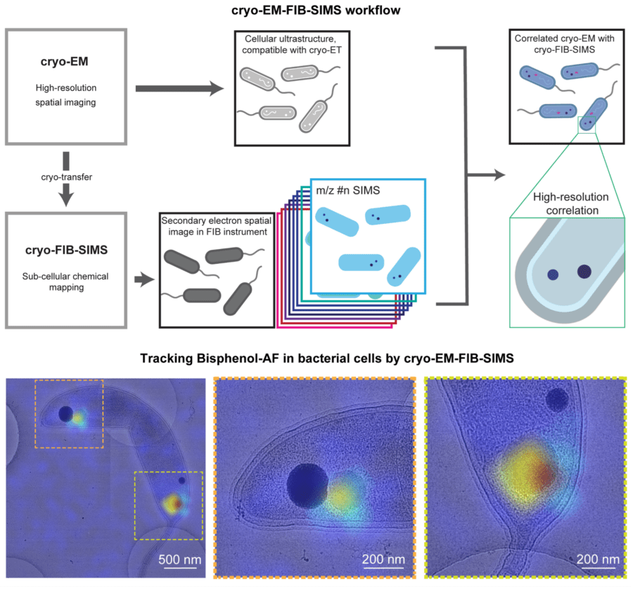
Specifically, the team examined the bioaccumulation of Bisphenol-AF (BPAF), a common chemical pollutant, in environmental bacteria. The team used the cryo-EM-FIB-SIMS workflow to visualise the subcellular localisation of BPAF, exploiting the technique’s strengths in determining spatial arrangement and chemical composition in cryogenic samples. BPAF was localised within large storage granules inside the bacteria, which raises questions regarding the possibility of its removal through the usual bacterial export machinery.
Looking ahead, this novel combination of spatial and chemical imaging provides a tool to investigate the interplay between the structure and function of molecules within cells, as well the interaction of molecules within their cellular environment, all at unprecedented levels of detail. The adaptability of the technique for different samples and imaging methods means it can be used to explore a wide range of biological processes, such as drug uptake by cells, uptake of a range of pollutants by bacteria and symbiotic metabolite sharing within microbiomes.
This work was funded by UKRI MRC, the Wellcome Trust and the Lister Institute for Preventative Medicine and EMBO.
Further references
Tanmay’s group page
Kiran Patil’s group – MRC Toxicology Unit
Electron Microscopy facility
Mass Spectrometry facility
Looking at Molecules: The electron cryomicroscopy revolution at the MRC LMB (video)
Related articles
A new method to prepare cryo-EM samples avoid protein damage during freezing
Gut microbes could protect us from toxic ‘forever chemicals’ – University of Cambridge