Key Advances
The LMB has remained at the forefront of molecular biology since our beginnings in the 1940s, pioneering new techniques and making world-changing discoveries about how life works. Here we highlight nine of these landmark scientific advances.
Explore our history timeline to see how further LMB ideas, discoveries and inventions have progressed science for over 60 years.
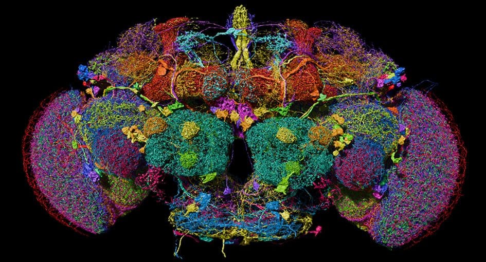
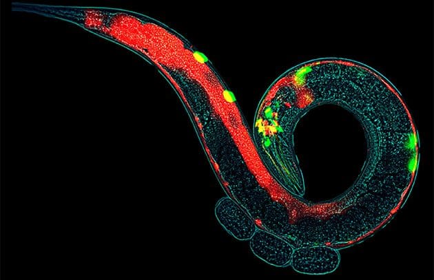
First complete ‘connectomes’
revealing the wiring map of brains
Just as a genome is a complete map of one organism’s DNA, so a connectome is a wiring map of a nervous system or brain.
By determining the first complete neuronal wiring diagrams of the nematode worm, fruit fly larva and adult, we are beginning to understand how memories are made and behaviour controlled.
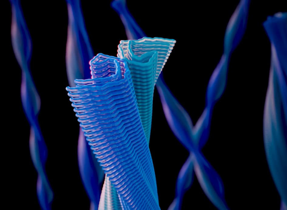
Structures of protein filaments in neurodegeneration
discovery of disease-related amyloids
The abnormal accumulation of protein amyloid filaments in brain cells defines human neurodegenerative diseases.
Cryo-EM has determined the molecular structures of major classes of neurodegeneration-linked filaments, revealing how specific structures are linked to different diseases.
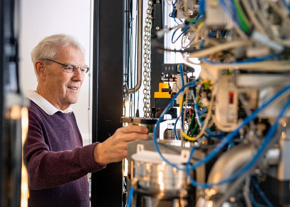
Electron cryomicroscopy (cryo‑EM)
visualising molecular machines in action
Imaging frozen biological samples using electrons has captured detailed snapshots of life’s most complex molecules performing their functions.
The 3D structures of these intricate molecular machines have vastly accelerated our understanding of how they work and is enabling the design of new drugs.
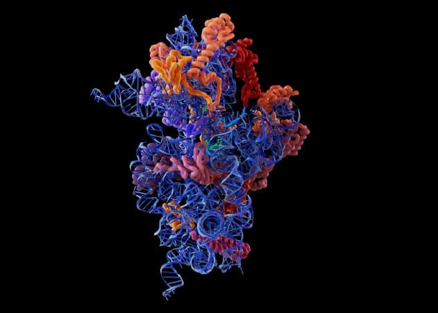
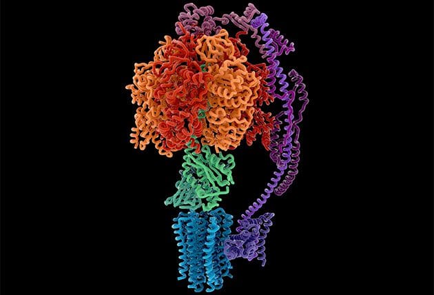
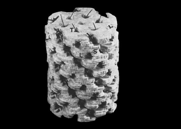
Structure of the ribosome
informing the design of new antibiotics
The ribosome carries out one of life’s core processes – translating the DNA code into proteins that carry out almost all biochemical reactions inside cells.
Solving the structure of this large molecular machine explained how it works and enabled the development of new antibiotics.
Structure of ATP synthase
revealing how energy is produced in cells
Solving the structure of ATP synthase revealed how this molecular machine uses an unprecedented rotary mechanism to produce ATP, a currency of energy used by all life.
Defects in this key enzyme are linked to a range of diseases, including heart failure, neurological dysfunction and cancer.
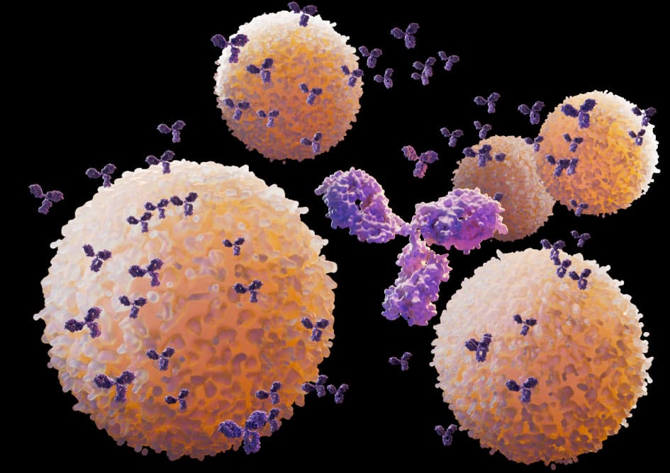
Monoclonal antibodies
providing targeted therapies for disease
Cells of the immune system were lab-engineered to indefinitely produce a single type, or monoclonal, antibody to target specific cells or molecules.
Monoclonal antibodies are now widely used to treat a range of chronic and severe conditions and are used in medical diagnostics such as pregnancy, HIV and blood group tests.
Control of cell growth
discovering the importance of programmed cell death
Mapping every cell division in the nematode worm, C. elegans, revealed that some cells are genetically programmed to die at defined points in development.
Preventing uncontrolled cell growth by targeting this cell death pathway is a key aim of cancer treatment. Conversely, the death of too many cells is responsible for the damage caused by strokes, heart attacks and Alzheimer’s disease.
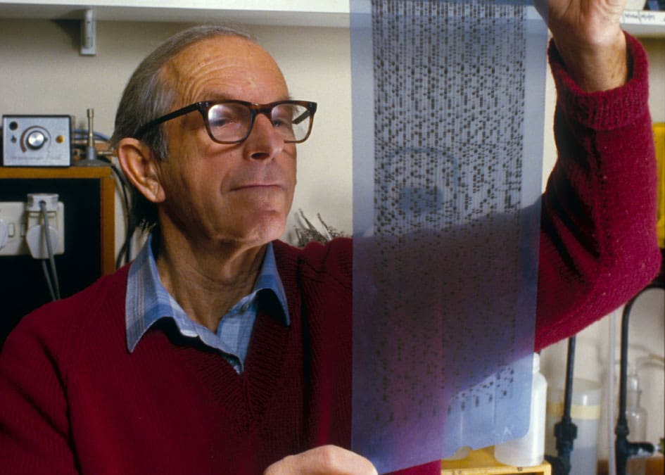
DNA sequencing
reading the code of life
The ability to read the exact sequence of the building blocks in DNA has transformed biology, modern medicine, forensics and agriculture.
The Sanger dideoxy method enabled the Human Genome Project, increasing understanding of genetic diseases and ushering in an era of personalised medicine.
Tomography
enabling 3D imaging in biology
The development of tomography, a method that reconstructs a 3D structure from a series of tilted 2D images, revolutionised much of biological and medical imaging.
For example, X-ray-based computed tomography (CT) scanning is now used routinely to produce 3D images for the diagnosis of neurological disorders, cancers and other diseases.
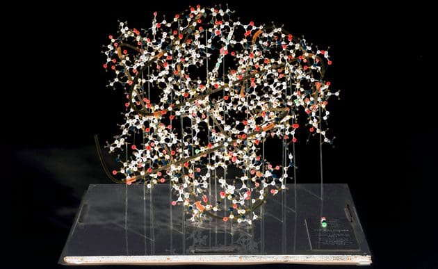
First atomic protein structure
enabling structure-based drug design*
Development of the X-ray crystallography technique led to the first 3D protein structures being solved: myoglobin and haemoglobin, the protein mutated in sickle cell anaemia.
These methods enabled the determination of structures for many proteins, revealing how they function, what goes wrong in disease and how they can be modulated with drugs.
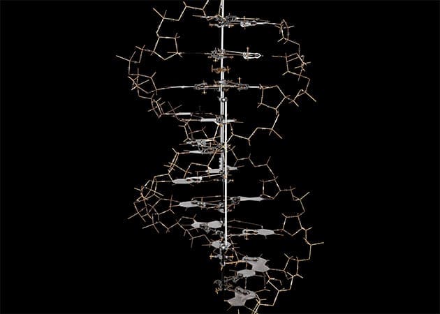
Structure of DNA
initiating the molecular biology revolution*
Solving the double-helical structure of DNA revealed how the hereditary code of life is stored, replicated and passed on to the next generation.
Understanding the structure of DNA opened the door to DNA sequencing, molecular genetics and biotechnology, underpinning modern biology and medicine.
* Research conducted in the MRC Unit for Research on the Molecular Structure of Biological Systems located within the University of Cambridge.