Long-overlooked cavities inside human sperm nuclei are revealed to be specialised compartments filled with protein-recycling machines
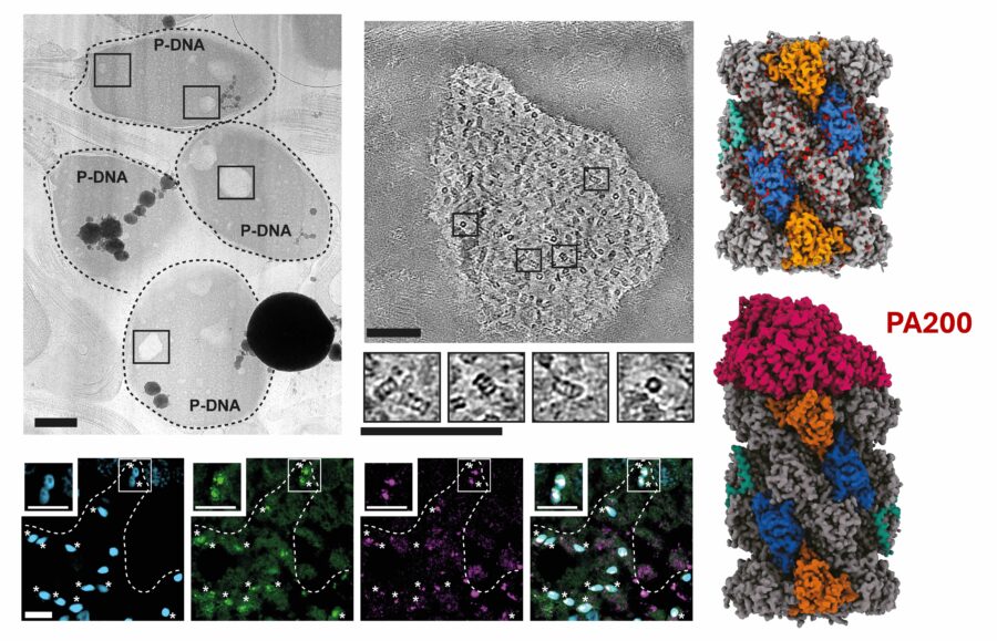
Proteasomes are vital, regulatory protein complexes which act as ‘protein recyclers’, breaking down proteins which are damaged, improperly folded or no longer required. They are essential for several cellular processes including spermatogenesis, the production of sperm cells. However, questions remain as to whether proteasomes are present in the sperm nucleus and, if so, where they are organised and how they operate. To answer this, Matteo Allegretti’s group in the LMB’s Structural Studies Division have used advanced microscopy to determine the molecular architecture of nuclear proteasomes, finding that they cluster in DNA-free cavities within the sperm nucleus.
The investigation was spearheaded by group members Piotr Kolata, Ália dos Santos and Tom Dendooven (who earlier this year launched his own independent research group at the VIB-VUB Center for Structural Biology). To probe the location and structure of proteasomes in human sperm cell nuclei, they began by using cryo-focussed ion beam milling to map human sperm cells, generating several incredibly thin, sheet-like layers named lamellae. Within the lamellae, the group noticed several small pockets which appeared lighter than the surrounding DNA. These cavities had been identified by microscopists many years ago, who named them lacunae, but it has remained unclear what they contain or whether they are biologically important.
The team used electron cryotomography (cryo-ET) to create detailed, 3D images of these lacunae, revealing that they are not merely empty spaces within the sperm nucleus. Instead, they contain large numbers of barrel-shaped protein complexes, which the researchers identified as proteasomes. To investigate whether this is unique to human sperm, the group repeated the imaging using mouse sperm cells. Mouse sperm were also found to contain proteasome-rich lacunae, but they were significantly smaller than the human counterpart.
Using electron cryomicroscopy (cryo-EM), the team determined the near-atomic structure of the nuclear proteasomes, revealing the presence of a component named a4s, which is largely unique to the testes and sperm cells. Interestingly, in one of the cryo-EM structures, the researchers found a small peptide fragment trapped inside an active site of the proteasome. This was an unexpected discovery, offering a snapshot of how the proteasome recognises and begins to break down proteins targeted for recycling.
Finally, the group studied samples of healthy human testes to identify when these proteasome clusters appear during spermatogenesis. They found that as sperm mature, proteasomes become increasingly concentrated in the nucleus. Clusters then begin appearing at the spermatid stage, when the head changes shape, the tail develops and excess cell material is discarded. Crucially, this is also the stage at which the DNA undergoes extreme compaction, going from being loosely wrapped around histones to tightly packed by protamines. The emergence of proteasome clusters to coincide with this suggests that the processes may be linked, with the proteasomes possibly removing excess proteins, including histones.
This research answers a fundamental question concerning how human sperm develop and prepare genetic material for the next generation. It sheds new light on the previously poorly understood lacunae cavities in sperm nuclei, revealing that these are proteasome-rich compartments which develop at a key point of spermatogenesis. While the presence of proteasome-rich lacunae is conserved across species, the work underlines species-specific differences in nuclear organisation, with lacunae appearing significantly larger in human sperm compared to mouse. More broadly, this work helps explain how sperm achieve one of the most remarkable feats in biology: compressing nearly two metres of DNA into a cell head just a few micrometres wide, while keeping that DNA functional for fertilisation. By determining the presence, organisation and function of proteasomes in the sperm nucleus, this study provides an anatomical and molecular framework for further studies to investigate the role of nuclear proteasomes in male fertility and reproductive health more widely.
This work was funded by UKRI MRC and the Wellcome Trust.
Further references
Matteo’s group page
The very first cell division – Meet Tom Dendooven (VIB blog)
Related articles
Hidden protein recycling hubs discovered inside human sperm cells (VIB news article)
Animal research statement
As a publicly funded research institute, the LMB is committed to engagement and transparency in all aspects of its research. This research used mice, in accordance with the UK Animals (Scientific Procedures) Act 1986. This work was conducted under a Project Licence, reviewed and approved by the MRC Laboratory of Molecular Biology (LMB) Animal Welfare and Ethical Review Body (AWERB) committee and the UK Home Office.
The LMB uses the minimum number of rodents necessary to achieve results and only uses animals in research where there are no suitable alternatives, in line with the 3R’s (replace, reduce, refine). We currently work with fruit flies, nematode worms, mice, rats and zebrafish.