A molecular cell atlas of the developing Drosophila nerve cord reveals a timestamp hidden within neurons which helps explain how complex circuits are built
The brain contains a vast number of neurons with specific shapes and molecular identities, organised into precise circuits. Much of this complexity is encoded in our DNA, ultimately determining the reproducible patterns of brain wiring that have been tuned by evolution to support the huge diversity of animal behaviour. But all neurons begin life looking rather similar: typically simple round cells originating from a neural stem cell. Given that we eventually observe such a wide variety of neurons, this begs the question, how does each neuron know its final shape and molecular identity? To answer this, Greg Jefferis’ group in the LMB’s Neurobiology Division have worked with former postdoctoral fellow, Erika Donà at the National Research Council (CNR) Institute of Neuroscience in Milan, Italy, and a group of international researchers. Spearheaded by LMB staff scientist Sebastian Cachero and Erika, the team have produced a cell atlas for the maturing fruit fly Ventral Nerve Cord (VNC, analogous to the spinal cord in vertebrates). This sheds new light on the construction of the nervous system, revealing a highly ordered process where a neuron’s birth order is the key determining factor to its identity.
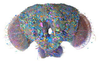
To assemble the atlas, the team profiled over 450,000 cells from the Drosophila VNC from male and female flies at four distinct stages of pupal development, covering the period where neurons establish their mature identities. The group then used molecular markers to distinguish cells based on key characteristics, including their stem cell of origin, or lineage. Crucially, to relate their molecular information to the shape and connections that exist in the mature VNC, the team integrated the new atlas with the complete connectome of the Drosophila VNC (another project from Greg’s group with collaborators at Janelia Research Campus and the University of Cambridge).
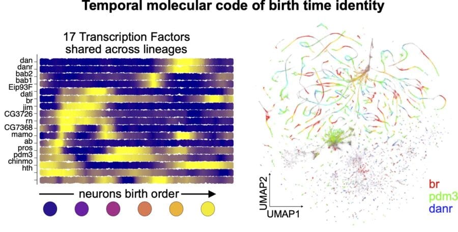
Their analyses revealed three organisational principles of the VNC which extend to the entire nervous system. First, the group discovered that neurons carry a molecular timestamp that records when they were born. This timestamp is encoded by 17 shared transcription factors, which together provide an identity code for birth order. Many of these transcription factors remain active into adulthood, highlighting that neurons carry this molecular record of birth date for a long time. As to how this impacts development, the group found that manipulating one of these transcription factors resulted in altered neuron gene expression and cell morphology, confirming that they are not just markers but can actively contribute to neuronal specification. Importantly, the team found that the same code is conserved across all neuronal lineages in the VNC, and extends to the central brain, suggesting it is a general principle used throughout most of the nervous system.
Secondly, the group observed that whether a neuron is born in the embryonic or larval stage of animal development is also crucial in determining molecular identity. During the embryonic stage of growth, the atlas highlights rapid diversification of neurons born subsequently from one progenitor, i.e. neuron’s molecular identities define clearly separated groups. Conversely, sibling neurons born during the larval stage diversify more gradually. This suggests that different developmental strategies are employed at different stages of life to guide diversification of neuronal types.
Collecting data from male and female flies, the group compared how neural development differs based on sex. Some neurons survive in males but undergo programmed cell death in females. Other neurons remain in both sexes but gradually, over the course of development, acquire different patterns of gene expression. This underlies the emergence of sex-specific circuitry that ultimately mediates sex-specific behaviours.
This atlas is a landmark resource for the field. By creating a detailed molecular map of neuronal development in Drosophila and linking it directly to the corresponding high-resolution connectome, the resulting material provides an unparalleled level of information to aid future studies investigating how complex nervous systems are built and how they operate in the adult. Birth order has long been linked to neuronal organisation, and this study demonstrates how this principle is enacted by a compact molecular code using just 17 transcription factors. This constitutes a beautiful example of how incredibly complex structures like the nervous system can be built from relatively simple instructions. Many of these transcription factors are conserved in vertebrates where timing mechanisms associated with early-born and late-born neurons have been reported. These findings likely reveal generalisable principles of nervous system development that may ultimately allow researchers to identify some of the processes that go wrong in developmental neurological disorders.
This work was funded by UKRI MRC, European Research Council, U.S. National Science Foundation NeuroNex, Wellcome, National Institutes of Health, Boehringer Ingelheim Fonds and the Cambridge Commonwealth, European and International Trust. It was further supported by the Blue Sky collaboration between AstraZeneca UK Limited and the Medical Research Council (BSF2-16), which supports pre-clinical research projects with the aim to improve understanding of fundamental biology and disease.
Further references
Greg’s group page
Erika Donà – CNR Institute of Neuroscience
Browse the VNC Atlas (requires account)
Related articles
Whole brain connectome of fruit fly is most complex brain ever mapped
Researchers reveal connectome of the male fruit fly central nervous system – Janelia