Human tau filaments serve as molecular templates for the assembly of identical mouse tau aggregates, explaining how neurodegenerative diseases spread across the brain
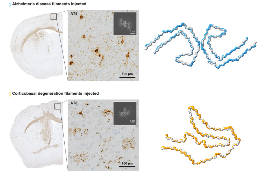
As neurodegenerative diseases such as Alzheimer’s progress, characteristic filamentous aggregates of abnormal tau proteins spread across the brain, causing symptoms to worsen. Exactly how these tau aggregates spread, whilst retaining their disease-specific structures, has been unclear. Continuing their long-established collaboration to investigate the molecular mechanisms behind neurodegeneration, Sjors Scheres and Michel Goedert’s groups, in the LMB’s Structural Studies and Neurobiology Divisions respectively, and Masato Hasegawa’s group at the Tokyo Metropolitan Institute of Medical Science (IGAKUKEN) have published new evidence showing that, akin to prion-caused neurodegeneration, misfolded human tau filaments act as a molecular template when injected in the mouse brain, causing typical mouse tau to adopt identical disease-specific folds.
The spread of misfolded filamentous aggregates of prion proteins, as occurs in diseases such as Mad Cow Disease (Bovine Spongiform Encephalopathy), Chronic Wasting Disease in deer or Creutzfeldt-Jakob disease in humans, is believed to be a form of self-propagation by templated seeding, whereby minute quantities of these filaments prompt native prion proteins to form filaments of the same structure. It has been theorised that misfolded tau filaments, which underpin over twenty different neurodegenerative conditions, may spread in the same manner. Previously, the Scheres, Goedert and Hasegawa groups have detailed how tauopathies can be characterised by their specific filament folds, meaning that the mechanism they use to spread across the brain must also conserve these unique structural features to advance the disease.
To investigate how this occurs, the group extracted abnormal tau aggregates from donated brains of people with either Alzheimer’s disease (AD) or corticobasal degeneration (CBD) and injected these aggregates into the brains of healthy mice. To track the progression of AD or CBD pathology, the mice were closely monitored over 12 months. Aki Shimozawa and Airi Tarutani at IGAKUKEN examined the mouse brains using several staining methods, including immunoblotting and immunoelectron microscopy, which established the spread of tau beyond the injection site and, importantly, that the pattern of spread differed for AD and CBD injected mice, mirroring how the disease progresses in humans.
This raised the question of whether the researchers were simply seeing human tau left over from the injections. To test this, the group used antibodies that distinguish human tau from mouse tau and examined filament extracts at several time points, including immediately after injection, one week later and after 12 months. The injected human tau disappeared quickly, and by 12 months, only mouse tau was detected, indicating that the later aggregates consisted of newly assembled mouse tau. This suggested that human tau had triggered a reaction from the mice’s own tau proteins to assemble into filaments.
For the final, crucial step in the project, the newly formed tau filaments from the mouse brains were extracted so that their atomic structures could be determined using electron cryomicroscopy (cryo-EM). Analysis of the resulting data by Sofia Lövestam, a postdoc in Sjors and Michel’s groups, revealed that mice injected with human AD tau developed mouse tau filaments with the characteristic AD fold. Likewise, mice injected with human CBD tau produced mouse tau filaments with the characteristic CBD fold. This established that the new aggregates in the mouse brains were structurally identical to the original human tau.
These results showed that, like prion diseases, tauopathies advance through the brain by using abnormal tau as a molecular template to produce more aggregates. This offers a mechanistic answer to why diseases like Alzheimer’s get progressively worse over the years. Crucially, the work adds credence to the group’s earlier argument that tauopathies should be classified based on their distinct folds, as the newly developed mouse tau remained faithful to the specific structures of the injected disease. These results underline how important biological information can be conveyed simply through protein shape. The spreading of tau aggregates through the brain requires cellular uptake of misfolded tau, copying the specific structure as well as the release and spread to neighbouring cells. Though the findings of this study don’t immediately offer a therapeutic answer to tau-based neurodegeneration, the mechanisms which control uptake, conversion and propagation each offer potential as a therapeutic target and should be further explored.
Finally, in showing that mouse-generated tau filaments are structurally indistinguishable from human tau filaments, the project affirms how important mouse models are for investigating neurodegenerative diseases. It is difficult for researchers to study how these diseases develop in human brains, and this work demonstrates how mouse models could be used to further investigate how tau spreads and, ultimately, how to stop it.
This work was funded by UKRI MRC, the Japan Agency for Medical Research and Development, the Japan Science and Technology Agency, the Japan Society for the Promotion of Science, Alzheimer’s Research UK and the Alzheimer’s Society.
Further references
Sjors’s group page
Michel’s group page
Masato Hasegawa – Tokyo Metropolitan Institute of Medical Science
Related articles
Classification of human tauopathies based on tau filament folds
Cryo-ET reveals new molecular insights into tau pathology in Alzheimer’s disease
New potential therapy for neurodegeneration removes tau aggregates and improves motor function
Animal research statement
As a publicly funded research institute, the LMB is committed to engagement and transparency in all aspects of its research. This research used mice, with all experimental protocols approved by the Animal Care and Use Committee of the Tokyo Metropolitan Institute of Medical Science.
The LMB only uses animals in research where there are no suitable alternatives and always uses the minimum number of rodents necessary to achieve results, in line with the 3R’s (replace, reduce, refine). We currently work with fruit flies, nematode worms, mice, rats and zebrafish.