Nobel Prize in Chemistry 1982
Aaron Klug
For his development of crystallographic electron microscopy and his elucidation of biologically important nucleic acid-protein complexes
“I like to think I would have achieved something elsewhere, but I could not have done what I have without so many able and gifted colleagues.”
Aaron Klug
Biological Structures in 3D
Electron microscopy has long been used to obtain two-dimensional (2D) pictures of biological objects. An electron microscope uses electrons to illuminate a specimen and create an enlarged image. They have much greater resolving power than light microscopes and can magnify specimens up to two million times, while the best light microscopes are limited to magnifications of 2,000 times. But unlike the light microscope, the electron microscope cannot be focused to view different levels: all the 3D matter in the line of view is projected into a 2D image.
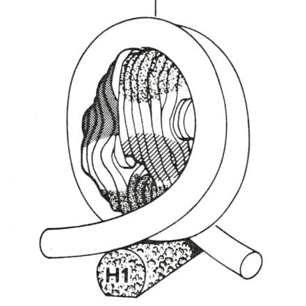
Aaron Klug overcame this limitation by taking images in different directions and combining them mathematically, using computers, to produce the 3D structure. He initially used this to determine the structure of viruses before studying the combination of protein and DNA in chromatin, of which chromosomes are made. Chromatin was broken into small fragments that could be examined. A model for chromatin was then proposed based on this knowledge of the structure of the fragments. The exact structure of chromatin affects how the genetic code along the DNA is read. This investigation was crucial in the understanding of cancer, in which the control of growth and division of cells by the genetic material no longer works.
Since the invention of 3D reconstruction, improvements in electron microscopes, specimen preparation and in computers have led to huge advances in 3D microscopy. Suitable specimens, such as viruses, can now be visualised in atomic detail. Sub-cellular structures can now be imaged by electron tomography, and similar approaches have revolutionised medical imaging, where computed tomography (CT) scanning is now used routinely to diagnose neurological diseases and cancers.