• Ivana Bukvin

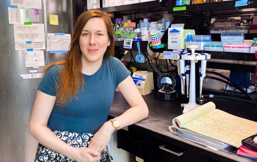
    The LMB is delighted to announce that Ivana Bukvin has been appointed as a Group Leader in the LMB’s PNAC Division. Beginning in January 2027, Ivana’s group will focus on co-translational folding and misfolding, with the ultimate goal of establishing a comprehensive structural and mechanistic understanding of how proteins fold during biosynthesis in cells.

    Commenting on her appointment, Ivana said, “How proteins fold in our cells has fascinated me for many years, and I think we are now reaching a really exciting point where advances in instrumentation and computing are beginning to make it possible to study this fundamental process in much greater depth and increasingly in the context of the cell itself. Being able to pursue this research at the LMB feels particularly fitting given its long tradition of supporting ambitious, long-term projects and its unique history in both ribosome biology and protein folding research. I am honoured and excited to start my lab at the LMB.”

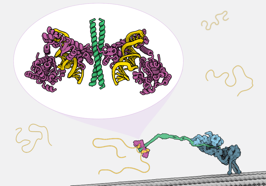
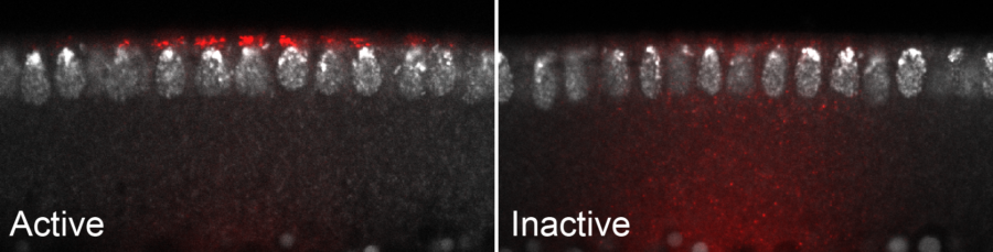
    Although protein folding is a fundamental cellular process, it is still unclear how folding, translation kinetics and interactions with ribosome-associated factors are coordinated within the crowded cellular environment to determine the fate of a newly synthesised protein. Ivana’s group will address this gap in knowledge, harnessing a novel platform to monitor co-translational folding and misfolding in real time.

    Specifically, Ivana seeks to utilise a single-molecule Förster Resonance Energy Transfer (smFRET) platform which works by using a photophysical mechanism to transfer energy from a donor molecule to an acceptor molecule, allowing for high-resolution, real-time analysis of nascent chain conformational changes and interactions. Using smFRET to monitor co-translational folding will allow Ivana to bridge structural detail, kinetic resolution and cellular context. Beyond gaining new insights in vitro, Ivana also aims to broaden application to in vivo studies, to better understand how protein folding happens in the more complex cellular environment.

    Whilst offering key insights into a significant process of life, Ivana’s research programme also holds wider clinical implications as aberrant translation and protein misfolding are defining characteristics of several diseases, including neurodegenerative diseases, which currently lack effective treatment. Therefore, knowing how and why protein folding and misfolding occurs is crucial to the future development of new interventions.

    Ivana began her career with a BSc in Biochemistry from the University of Belgrade, Serbia. She followed this with an MSc at University of Tübingen, Germany, during which she completed part of her thesis work as a visiting scholar at University College London, where she first began investigating co-translational folding, developing a force-based translational assay to monitor the process in vitro and in cellulo. She later received her PhD from University College London in 2023, where she worked with John Christodoulou to establish biochemical and NMR methods to investigate ribosome-nascent chain complexes. Since then, Ivana has been a postdoctoral scholar at Stanford University, USA, where she has worked with Judith Frydman to investigate the role of translation dynamics in Huntington’s disease. With the support of the Huntington’s Disease Foundation postdoctoral fellowship, she has begun establishing the smFRET approach to characterise the conformational landscape of nascent huntingtin on the ribosome.

    Ivana’s recruitment to the LMB was supported by the Global Talent Fund, a £54 million government investment, shared between 12 UK organisations including the LMB, designed to attract leading researchers and their teams to the UK.

    Further references

    Ivana’s group page
    Protein and Nucleic Acid Chemistry (PNAC) Division

  • A novel degradative pathway, antibody-directed xenophagy, protects cells from viral and bacterial infection

    Timelapse movie of TRIM21 spots (cyan) co-localising with cell-invading adenoviruses (magenta). Captured by Anna Albecka and Tyler Rhinesmith.

    In order to prevent infection, mammals have evolved multiple complementary antimicrobial factors which integrate into sophisticated and robust immune systems. One important factor in human immunity is TRIM21, an intracellular protein which binds to antibodies. TRIM21 is an E3 ubiquitin ligase, an enzyme which catalyses ubiquitination, the transfer of the signalling protein ubiquitin onto ‘client’ molecules. Previously, Leo James’s group described how TRIM21 protects from viral infection by binding to antibody-coated viruses in the cytosol of cells, triggering the viruses to be ubiquitinated and degraded. However, the mechanism the cell uses to degrade these intercepted viruses remained elusive.

    In a new study led by Tyler Rhinesmith, researchers have now discovered a novel pathway called antibody-directed xenophagy (ADX) which explains how TRIM21 protects cells from infection by diverse pathogens. A postdoctoral scientist in the Leo’s group, Tyler conducted a genome-wide CRISPR/Cas9 knockout screen, individually removing every gene across the human genome and testing how its deletion impacted TRIM21-triggered degradation of viruses. The results were striking, revealing a previously undescribed process by which TRIM21 is able to trigger autophagy of cell-invading viruses.

    Autophagy, literally ‘self-eating’, is a conserved cellular process through which the cell delivers damaged or toxic components to acidic organelles for degradation and recycling. While autophagy is crucial for the maintenance of cellular health, its ability to defend against viral infection has not been very well studied. So, the team dissected the ADX pathway in molecular detail.

    In order to do this, Anna Albecka, a staff scientist in the Leo’s group, developed a high-fidelity confocal microscopy platform which allowed the team to visualise previously unidentified events in the TRIM21 restriction mechanism. For the first time, the researchers could observe binding of TRIM21 to antibody-coated viruses inside cells, in real time. After TRIM21 ubiquitinates the invading virus complex, Anna’s microscopy demonstrated that ubiquitin stimulates the assembly of autophagy components around viruses, including LC3, a marker for membranous compartments called autophagosomes.

    Super-resolution image of an LC3-positive autophagosome engulfing a TRIM21- and antibody-coated adenovirus. Credits Claudia Puri, Matthew J. Gratian, Anna Albecka, and Tyler Rhinesmith.

    Working with Claudia Puri and David C. Rubinsztein at the UK Dementia Research Institute Cambridge, the team used super-resolution microscopy to visualise the assembly of these autophagosome membranes around individual viral particles coated in antibodies and TRIM21. Together these observations revealed the step-wise process by which incoming virions are incarcerated inside sealed, LC3-positive autophagosomes. Anna was further able to show that these virus-containing autophagosomes are ultimately delivered to acidic lysosomes, resulting in the degradation of each virus into harmless peptides and nucleotides. Significantly, this study shows that antiviral autophagy is a highly effective strategy deployed by cells to protect themselves from infection and provides new tools for investigating this process.

    Inspired by the ability for TRIM21 to activate by clustering around clients of very different architectures, the team next sought to understand whether it could also intercept a completely different type of pathogen: bacteria. The team used antibodies and a novel live cell microscopy method to track bacterial growth inside mouse cells. They observed the same ADX pathway that intercepts viral infection also potently restricts growth of intracellular Salmonella. This discovery is significant because it explains how TRIM21 is able to intercept and trigger degradation of invading pathogens of many complex structures and diverse lineages.

    By leveraging the intrinsic flexibility of the autophagy pathway, ADX can adapt to and degrade variety of large and difficult targets. This demonstrates that the cell does not require a bespoke defence strategy for every individual pathogen. Instead, it employs a universal strategy, reliant on TRIM21, to redirect the cell’s existing autophagy machinery to any harmful material tagged with antibodies. This adaptability makes ADX clinically important for human immunity and, excitingly, a potential target for therapeutic enhancement.

    This work was funded by UKRI MRC, the UK Dementia Research Institute, the Wellcome Trust, and the Cambridge Trust.

    Further references

    TRIM21 induces selective autophagy of viruses and bacteriaRhinesmith T, Albecka A, Vaysburd M, Puri C, Luptak J, Boulanger J, Nguyen Le QM, Gratian MJ, O’Connell K, Few L, Donaldson-Wing M, Kozik P, Rubinsztein DC, James LCMolecular Cell: (2026)

    Leo’s group page
    Claudia Puri – Cambridge Institute for Medical Research
    David Rubinsztein – Cambridge Institute for Medical Research

    Related articles

    New potential therapy for neurodegeneration removes tau aggregates and improves motor function
    Understanding the protein modifications behind Trim-Away protein degradation technology
    Classification of human tauopathies based on tau filament folds

    Animal research statement

    As a publicly funded research institute, the LMB is committed to engagement and transparency in all aspects of its research. This research used mice, in accordance with the UK Animals (Scientific Procedures) Act 1986. This work was conducted under a Project Licence, reviewed and approved by the MRC Laboratory of Molecular Biology (LMB) Animal Welfare and Ethical Review Body (AWERB) committee and the UK Home Office. 

    The LMB uses the minimum number of rodents necessary to achieve results and only uses animals in research where there are no suitable alternatives, in line with the 3R’s (replace, reduce, refine). We currently work with fruit flies, nematode worms, mice, rats and zebrafish.

    More on how the LMB uses animals in research.

  • Combining cryo-EM and mass spectrometry imaging capabilities into a single workflow provides unprecedented spatial and chemical composition detail of biological samples

    Imaging mass spectrometry correlated with cryo-EM yields insights into subcellular localization of elements and molecules.

    Electron cryomicroscopy (cryo-EM) is an invaluable tool for determining molecular structures, capable of visualising biological samples with high spatial resolution. However, whilst the technique allows researchers to distinguish shapes and contrast of features in their images, it cannot provide any information regarding the chemical composition of the imaged sample. Conversely, mass spectrometry is a technique which provides compositional information but is unable to give detailed spatial information. Akin to adding colour to a black-and-white photograph, augmenting cryo-EM spatial data with mass spectrometry-derived chemical detail would greatly expand the information scientists can gather. Tanmay Bharat’s group, in the LMB’s Structural Studies Division, has developed a novel workflow for correlated electron microscopy and imaging mass spectrometry which achieves just this.

    Spearheaded by postdoc Hannah Ochner, the new workflow first captures high resolution structural information using cryo-EM, followed by imaging mass spectrometry of the same sample using a focussed ion beam scanning electron microscope adapted with a time-of-flight mass spectrometer (FIB-SIMS, focussed ion beam secondary ion mass spectrometry), allowing correlation between both types of data. Using this new cryo-EM-FIB-SIMS workflow, the team were able to track compounds at unprecedented resolutions, providing a subcellular map with more information than either of the composite techniques can singularly.

    This new workflow is compatible with numerous sample types and can be combined with other imaging methods. Using dual metal and fluorescently tagged bacteria prepared by Buse Isbilir, another postdoc in the group, with PhD student Yuexuan Zhang, the team integrated cryo-EM-FIB-SIMS with cryo-light microscopy, enabling investigation of samples with three imaging modalities. The new workflow can also be combined with electron cryo-tomography (cryo-ET) of FIB-milled ultra-thin sections (lamellae) of multicellular samples. This versatility means it could be applied to a wide range of biological and biomedical investigations.

    To demonstrate just one avenue of its use, Tanmay’s group collaborated with Kiran Patil’s group at the MRC Toxicology Unit to examine how chemical compounds such as fluorinated pollutants bioaccumulate in environmental bacteria. These pollutants, commonly referred to as ‘forever chemicals’, are one of the most urgent public health problems requiring research to better understand the risks associated with microplastics and pollution, and to identify novel solutions.

    Correlated cryogenic electron and mass spectrometry imaging used to study the uptake of microplastics pollutants by environmental bacteria.

    Specifically, the team examined the bioaccumulation of Bisphenol-AF (BPAF), a common chemical pollutant, in environmental bacteria. The team used the cryo-EM-FIB-SIMS workflow to visualise the subcellular localisation of BPAF, exploiting the technique’s strengths in determining spatial arrangement and chemical composition in cryogenic samples. BPAF was localised within large storage granules inside the bacteria, which raises questions regarding the possibility of its removal through the usual bacterial export machinery.

    Looking ahead, this novel combination of spatial and chemical imaging provides a tool to investigate the interplay between the structure and function of molecules within cells, as well the interaction of molecules within their cellular environment, all at unprecedented levels of detail. The adaptability of the technique for different samples and imaging methods means it can be used to explore a wide range of biological processes, such as drug uptake by cells, uptake of a range of pollutants by bacteria and symbiotic metabolite sharing within microbiomes.

    This work was funded by UKRI MRC, the Wellcome Trust and the Lister Institute for Preventative Medicine and EMBO.

    Further references

    Subcellular chemical mapping using correlated cryogenic electron and mass spectrometry imaging.Ochner H, Isbilir B, Blasche S, Scheidweiler D, Zhang Y, Wang Z, Smith T, Franco C, Bradley R, Patil KR, Bharat TAMNat Methods

    Tanmay’s group page
    Kiran Patil’s group – MRC Toxicology Unit
    Electron Microscopy facility
    Mass Spectrometry facility
    Looking at Molecules: The electron cryomicroscopy revolution at the MRC LMB (video)

    Related articles

    A new method to prepare cryo-EM samples avoid protein damage during freezing
    Gut microbes could protect us from toxic ‘forever chemicals’ – University of Cambridge

  • White mouse held gently in purple-gloved hands, with a green lab coat in the background.

    The LMB has been granted Leaders in Openness status for 2026-2029. This UK-wide recognition is awarded to organisations demonstrating sector-leading transparency in communications concerning the use of animals in research. It builds upon the Concordat on Openness on Animal Research in the UK, established in 2014, to encourage open and honest public dialogue.

    First introduced in 2019, Leaders in Openness status is administered by Understanding Animal Research, a not-for-profit organisation dedicated to advancing societal comprehension of the humane use of animals in research in the UK. It is given only after completion of a rigorous application process, assessed by public representatives and peer reviewers, in which applicants must demonstrate how they have gone above and beyond to make the principles of openness and transparency central to their operations. The status is awarded for three years, after which holders must reapply, providing evidence of their continued leadership and dedication to explaining when, how and why animals are used in research.

    This recognition is a significant milestone for the LMB. The LMB’s Biological Services facility carefully manages breeding and experimental colonies of mice and rats, and animal welfare is central to their work. Rigorous oversight, regular assessments and specialist training ensure animals are used only where no suitable alternatives exist and that the highest standards of care are upheld. In parallel, the LMB actively pursues and develops replacement approaches wherever possible. The assessment framework for Leaders in Openness status has provided clear guidance for the LMB to follow to be more open and visible about this work and the standards that underpin it.

    As part of this commitment to openness, the LMB has worked to make information about its use of animals clear, accessible and easy to find. This includes publishing detailed statistics on the numbers of animals used and the types of experiments conducted each year, alongside explanations of why this work is necessary. The LMB also shares this information through its social media channels, including videos illustrating measures implemented to further the principles of Replacement, Refinement and Reduction (the 3Rs) and a virtual tour of the animal facility. Research articles that involve work with mice or rats are clearly labelled online so readers can understand how the research was carried out.

    Beyond digital communications, LMB animal technicians have led in‑person engagement, developing a rodent care activity for the 2023 LMB Open Day, which attracted more than 2,500 visitors, hosting a bespoke friends‑and‑family visit to the animal facility attended by over 50 people and delivering workshops and animal facility tours for work experience students. This sustained approach to transparency has previously been recognised with an Openness Award from Understanding Animal Research.

    Lesley Drynan, Head of Biological Services, commented: “We are delighted that the LMB has been recognised by UAR as a Leader in Openness. This achievement reflects the ongoing commitment and dedication of LMB staff to transparency in our use of animals in research, to maintaining the highest standards of animal welfare and to upholding scientific integrity. We are proud to receive this recognition, which highlights the progress we have made on our openness journey. We look forward to continuing this in collaboration with UAR, whose support and partnership have been instrumental in enabling this progress.”

    Further references

    Leaders in Openness 2026-2029 – Understanding Animal Research
    LMB Animal Research
    LMB wins Openness Award from Understanding Animal Research

  • Discovery of a code that allows molecular motors to select specific mRNAs for subcellular localisation

    Structural representation of the Egl-BicD-dynein-dynactin complex transporting an mRNA molecule along a microtubule, with a magnified inset of the Egl-BicD-RNA structure.
    Structural model of the dynein-adaptor complex transporting an mRNA along a microtubule and, inset, a magnified view of the RNA-bound adaptor complex.

    In order to perform their elaborate functions, cells must position the right proteins in the right place at the right time. In many cases, this spatial organisation is achieved by localising the instructions for making proteins – mRNA molecules – to specific sites, thereby ensuring their products are only made and function where needed. It has been recognised for decades that mRNA delivery is mediated by large machines called molecular motors, which translocate along polarised cytoskeletal tracks together with mRNA cargo. However, it has not been clear how motors select specific mRNAs for transport from a much wider pool of these molecules. This is because there are no obvious similarities in the sequences or predicted structures of different localising mRNAs.

    A collaborative project between the groups of Simon Bullock and Andrew Carter, in the LMB’s Cell Biology and Structural Studies Divisions, respectively, has revealed that mRNAs are chosen by motor complexes based on a set of shared features that had previously gone unnoticed.

    Using electron cryomicroscopy, Kashish Singh, a postdoc in Andrew’s group, was able to visualise the structures of several mRNAs bound by an adaptor protein complex for the microtubule-based motor dynein.

    The RNA-binding function of the adaptor complex is provided by a protein called Egalitarian (Egl), which was found to engage targets by assembling multiple, atypical RNA-binding domains around double-stranded RNA stem loops. The structures also revealed that the Egl-binding region in different RNAs adopts a highly similar three-dimensional fold, despite being formed by very different sequences. By studying how mutations that disrupt this fold affect binding to Egl, as well as dynein-mediated mRNA transport in fruit fly embryos, Sabila Chilaeva, a PhD student in Simon’s group, demonstrated that the shape of the stem loop is critical for mRNA recognition.

    Images of blastoderm-stage Drosophila embryos with either localised or non-localised distributions of fluorescent mRNAs. mRNA signals are in red and nuclei are in grey
    An mRNA containing RNA elements that conform to the rules of recognition by Egl is transported to the apical side in the fruit fly embryo by dynein (left), whereas one lacking these features remains in the basal cytoplasm (right). mRNAs, red; nuclei, grey.

    The team found that association with Egl also depends on the identities of two specific base pairs in the RNA structures, which are spaced a set distance apart in their helical regions. These features are discriminated by side chains of two structurally related domains in the protein, which reach into the RNA’s minor groove. This level of sequence specificity has not previously been demonstrated for proteins that bind double-stranded RNA, expanding our understanding of how molecular recognition can be achieved in RNA biology.

    Reinforcing the importance of the identified structural and sequence features, grafting them into an otherwise inactive RNA stem loop was sufficient to generate a fully active localisation signal.

    Additional experiments, including reconstitution of mRNA-motor complexes with purified components by Mark McClintock, a senior investigator scientist in Simon’s group, revealed an additional selection criterion: the presence of two Egl-binding structures on the same mRNA molecule. This constitutes a quality control mechanism that ensures only bona fide mRNA cargo is moved by the transport machinery, thereby preventing the potentially damaging accumulation of proteins at ectopic cellular sites.

    Collectively, the project has revealed that motors select mRNAs for intracellular transport based on the shape and sequence of individual RNA elements, as well as the presence of multiple analogous signals in the same molecule. By providing a molecular explanation for how diverse localising mRNAs are selected, this work resolves a long-standing question in cytoskeletal transport. It also suggests that similar recognition principles may operate in other processes governed by RNA-binding proteins, including translational control, mRNA decay and RNA silencing.

    This work was funded by UKRI MRC, UKRI BBSRC and EMBO.

    Further references

    Structural basis for recognition of diverse localizing mRNAs by Egl–BicDSingh K, Chilaeva S, McClintock MA, Carter AP, Bullock SLNature Structural & Molecular Biology 33(5): 882-893 (2026)

    Simon’s group page
    Andrew’s group page

    Related articles

    Cracking the (zip)code of dynein-dependent RNA localizationBesse F, Ramos ANature Structural & Molecular Biology 33(5): 739-741 (2026)

    A genome-wide resource for dissecting transport within cells
    Cryo-EM reveals first high-resolution structure of the dynein-dynactin complex bound to microtubules
    Packaging molecular motors for delivery

  • Edith Heard
    Image courtesy of the Francis Crick Institute

    The LMB is thrilled to welcome Edith Heard, CEO of the Francis Crick Institute, to deliver the 2026 César Milstein Lecture. Edith’s talk, titled ‘Life with two X chromosomes: mechanisms of silencing and escape during X-chromosome inactivation’ will begin at 11am (BST) on the 11th May 2026. External attendees are advised to arrive at least ten minutes in advance to allow time to register at LMB Reception.

    Edith Heard joined the Francis Crick Institute in 2025. In addition to serving as Chief Executive Officer, she leads a research group focussed on developmental epigenetics. More specifically, she uses multi-disciplinary approaches to investigate how X-chromosome inactivation is initiated and maintained in developing female embryos.

    She is responsible for several breakthroughs in this field, including uncovering how the RNA molecule Xist silences one of the X chromosomes. Her work has also identified the protein complexes and chromatin architecture which underpins this process, highlighting key developmental timing, plasticity and epigenetic reprogramming events which occur in early embryogenesis. More recently, her group has shown that modifying Xist levels causes inactivation of genes which normally escape X chromosome silencing, extending into later stages of development, illustrating that Xist is not only important at the very early stages of embryo growth.

    Edith began her career in science at the University of Cambridge, where her undergraduate studies specialised in genetics. After graduating in 1986, she pursued a PhD investigating gene amplification mechanisms in the context of cancer at the Imperial Cancer Research Fund before turning her attention to X-chromosome inactivation as a postdoc at the Pasteur Institute in Paris. She later spent a year as a visiting scientist at Cold Spring Harbor Laboratory, USA, before she joined the Institut Curie, France as Director of the Department for Genetics and Developmental Biology. In 2019, she was appointed Directed General of EMBL and oversaw operations across its six sites during the Covid-19 pandemic.

    Edith became a member of EMBO in 2005 and was elected a Fellow of the Royal Society in 2013. Her work has been recognised with the CNRS Silver Medal in 2008, the CNRS Gold Medal in 2024 and the Croonian Medal and Lecture from the Royal Society in 2025.

    Lecture abstract

    Dosage compensation is essential to balance gene expression between the sexes, as females carry two X chromosomes while males carry only one. In mammals, this balance is achieved through X-chromosome inactivation, a process by which one X chromosome is transcriptionally silenced in female cells to prevent a potentially deleterious double dose of X-linked gene products. X inactivation has long been viewed as a stable and irreversible mechanism established early in development and faithfully maintained throughout life. However, it is now clear that this process is far more dynamic than previously thought. During early embryogenesis, X inactivation can be reversed, resetting epigenetic states before being re-established in a lineage-specific manner. Moreover, a substantial number of genes can escape silencing in somatic tissues, leading to variable expression from the inactive X chromosome.

    My group has contributed to this revised paradigm by investigating the extent and regulation of escape from X inactivation, and by demonstrating the transient nature of silencing during early development. My talk will focus on the molecular mechanisms that underlie both gene silencing and escape, including the roles of chromatin organization, long non-coding RNAs and nuclear architecture. I will highlight how these findings contribute to our understanding of epigenetic stability and plasticity, with implications for sexual dimorphism and disease.

    Background information

    The César Milstein Lecture is named in honour of César Milstein, an LMB Nobel Laureate. This named lecture is one of a series of named lectures organised by the LMB and given by eminent scientists from around the world. These talks are supported financially by AstraZeneca and the Max Perutz Fund.

    César was born in Argentina in 1927. After completing PhDs in both Buenos Aires and Cambridge, he embarked on a brief spell of research in Argentina before he joined the LMB in 1963. César then spent the rest of his career and his life here.

    César developed an early interest in immunology, with his research concentrated on antibody structure and diversity. In the early 1970s, he and his postdoc, Georges Köhler, developed the technique used to produce monoclonal antibodies. This work led to them being jointly awarded the 1984 Nobel Prize in Physiology or Medicine. The technique developed by César and Georges has since been developed further by LMB colleagues for therapeutic applications, leading to the creation of several MRC spin-out companies. César continued his research on how somatic mutation arises in immunoglobulin genes. He died in Cambridge on 24th March 2002.

    Further references

    Edith Heard’s group page
    César Milstein
    LMB Named Lectures

Loading...