Group Leader

Lalita Ramakrishnan

Mechanisms of tuberculosis pathogenesis

Lalita Ramakrishnan
Group Members
  • Lubos Chvostek
  • Ana de Oliveira
  • Michal Eisenberg‑Bord
  • Harry Fisher
  • Adam Fountain
  • Nicola Goodwin
  • Jonathan Hill
  • Kenny Jungfer
  • Meindert Lamers
  • Eloise Mawdesley
  • Joana Milão
  • Michael Price
  • Damian Quigley
  • Jonathan Shanahan
  • Katie Shanahan
  • Nathan Usher
  • Laura Whitworth
  • Lucinda Wood

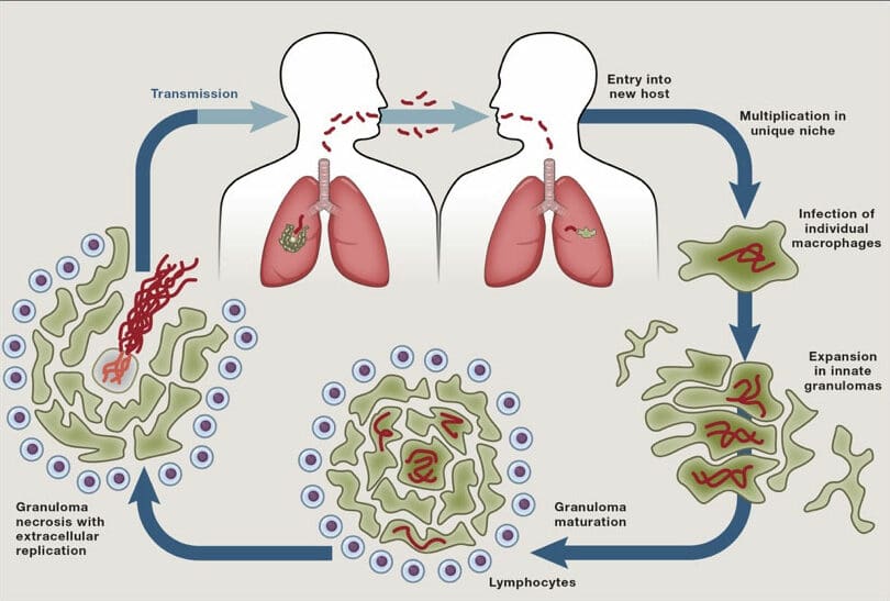
Tuberculosis (TB) remains a major cause of death despite the existence of a live attenuated vaccine (BCG) for a century and effective antibiotics for 60 years. TB’s persistence over millennia in the face of these major medical advances underscores the ability of Mycobacterium tuberculosis to evade and exploit host defences and antibiotics. Pathogenic mycobacteria infect macrophages and survive in these primary immune defence cells by subverting their endocytic trafficking and microbiocidal mechanisms. The bacteria also alter macrophage differentiation, migration and aggregation to induce the formation of granulomas, the enigmatic, complex, organised immune structures that can paradoxically promote bacterial growth. Ultimately, mycobacteria can cause granuloma breakdown by inducing programmed macrophage death, a step that further increases bacterial growth and promotes transmission to new hosts.

Coloured flow diagram showing cycle of Mtb entry into human host, developing granuloma, breakdown of the granuloma and transmission to a new human host.
M. tuberculosis ‘life cycle’ (from Cambier et al., Cell 2024).

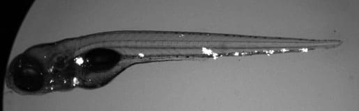
We use zebrafish larvae as a surrogate model organism to study TB, exploiting its optical transparency and genetic and pharmacological tractability to monitor infection in real time. Through genetic screens, we identify host susceptibility and resistance factors and bacterial virulence determinants that alter specific infection steps. Our research is shedding light on TB pathogenesis as well as fundamental macrophage biology – organellar cross-talk, migration, adhesion and death. Our findings have been borne out in humans and are informing new treatment strategies. We have also used the zebrafish to unravel mysteries in other granulomatous diseases like leprosy and schistosomiasis.

Black and white image of a translucent zebrafish larva with white areas indicating the presence of fluorescent mycobacteria Caption text
Zebrafish larva infected with fluorescent mycobacteria.

Selected Publications

mTOR-regulated mitochondrial metabolism limits mycobacterium-induced cytotoxicity.Pagán AJ, Lee LJ, Edwards-Hicks J, Moens CB, Tobin DM, Busch-Nentwich EM, Pearce EL, Ramakrishnan LCell 185(20): 3720-3738.e13 (2022)
Tumor necrosis factor induces pathogenic mitochondrial ROS in tuberculosis through reverse electron transport.Roca FJ, Whitworth LJ, Prag HA, Murphy MP, Ramakrishnan LScience 376(6600): eabh2841 (2022)
TNF Induces Pathogenic Programmed Macrophage Necrosis in Tuberculosis through a Mitochondrial-Lysosomal-Endoplasmic Reticulum Circuit.Roca FJ, Whitworth LJ, Redmond S, Jones AA, Ramakrishnan LCell 178(6): 1344-1361.e11 (2020)
A Macrophage Response to Mycobacterium leprae Phenolic Glycolipid Initiates Nerve Damage in Leprosy.Madigan CA, Cambier CJ, Kelly-Scumpia KM, Scumpia PO, Cheng TY, Zailaa J, Bloom BR, Moody DB, Smale ST, Sagasti A, Modlin RL, Ramakrishnan LCell 170(5): 973-985.e10 (2017)